ECG Heart Block
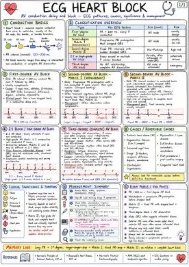
Conduction Basics: - Heart block = impaired impulse conduction from atria to ventricles, usually at the AV node, His bundle, or bundle branches. - PR interval (normal): 120–200 ms. - AV block severity ranges from delay → intermittent non-conduction → complete AV dissociation. Classification Overview: - First-degree AV block: PR > 200 ms, every P conducted - Second-degree Mobitz I (Wenckebach): Progressive PR prolongation then dropped QRS - Second-degree Mobitz II: Fixed PR intervals with sudden dropped QRS - 2:1 or high-grade AV block: Every second or multiple P waves blocked - Third-degree AV block: No AV relationship, complete AV dissociation First-Degree AV Block: - ECG: PR interval > 200 ms, constant PR, each P followed by QRS - Usually asymptomatic - Causes: ↑ vagal tone, athletes, β-blockers, non-DHP CCBs (verapamil, diltiazem), digoxin, ischemia, myocarditis - Clinical pearl: Not a true dropped beat; it is conduction delay only Second-Degree AV Block - Mobitz I (Wenckebach): - ECG: Progressive PR prolongation until a non-conducted P wave occurs; then cycle repeats (Grouped beating) - Usually nodal - Often transient; vagotonia, inferior MI, drugs - Usually less likely to progress to complete heart block than Mobitz II - Management: Often observation unless symptomatic Second-Degree AV Block - Mobitz II: - ECG: PR interval constant in conducted beats with intermittent sudden dropped QRS - Usually infranodal (His-Purkinje); often associated with wide QRS - Important causes: anterior MI, fibrosis/sclerosis of conduction system, myocarditis, infiltrative disease - High risk of progression to complete heart block and syncope - Management pearl: Treat seriously; pacing usually indicated 2:1 Block / High-Grade AV Block / 2:1 Block: - 2:1 AV block: Every alternate P wave not conducted - High-grade AV block: ≥ 2 consecutive non-conducted P waves - Distinction between Mobitz I and II may be difficult in 2:1 block - Clues favoring infranodal disease: wide QRS, very low ventricular rate, structural heart disease - Emphasize careful monitoring and pacing consideration Third-Degree (Complete) AV Block: - ECG: Complete AV dissociation; P waves and QRS complexes occur independently - Atrial rate > ventricular escape rate - Junctional escape rhythm: often narrow QRS and 40–60/min - Ventricular escape rhythm: often broad QRS and 20–40/min - Symptoms: fatigue, dizziness, syncope (Stokes-Adams), hypotension, heart failure - Causes: ischemia/infarction, degeneration, post-cardiac surgery, congenital block, Lyme carditis, hyperkalemia, drug toxicity - Emergency note: Unstable patient requires immediate pacing Causes / Reversible Causes: - Ischemic heart disease / MI - Degenerative fibrosis (Lev / Lenegre) - Increased vagal tone - Drugs: β-blockers, verapamil, diltiazem, digoxin, amiodarone - Electrolytes: hyperkalemia - Myocarditis / Lyme disease - Infiltrative disease: sarcoidosis, amyloidosis - Post-procedural / post-cardiac surgery - Congenital heart block Clinical Significance & Symptoms: - None - Fatigue - Dizziness / Presyncope - Syncope (Stokes-Adams) - Dyspnea - Chest pain - Hypotension - Symptoms range from none to fatigue, presyncope, syncope, dyspnea, chest pain, hypotension - Severity depends on level of block, escape rhythm reliability, and underlying cardiac disease - Mobitz II, high-grade AV block, and complete heart block are clinically important because they may cause low cardiac output and sudden deterioration Management Summary: - Assess ABCs, vitals, symptoms, hemodynamic stability - Stop AV nodal blocking drugs if appropriate - Check ECG, electrolytes, troponin, drug history, ischemia/infection clues - Symptomatic bradycardia: Atropine 0.5 mg IV q3–5 min (max 3 mg) – may help if nodal block; usually ineffective in infranodal block - If unstable (hypotension, altered mental status, ischemia, shock, HF): Transcutaneous pacing immediately; Transvenous pacing as needed - Permanent pacemaker usually indicated for Mobitz II, high-grade AV block, or complete heart block not due to reversible cause - Treat the underlying cause Exam Pearls / Viva Points: - PR > 200 ms = first-degree AV block - Wenckebach = progressive PR prolongation before dropped beat - Mobitz II = fixed PR with dropped beat = dangerous - Third-degree block = AV dissociation - Wide QRS often suggests infranodal disease - Inferior MI more often causes nodal block; anterior MI more often causes infranodal block - Atropine may help nodal block; usually ineffective in infranodal block - Always look for reversible causes - Pacing indicated in symptomatic patients or high-risk blocks