
Cardiac Anatomy and Function
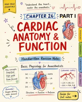
Cardiac Anatomy & Function: - Oxygen delivery meets demand - Structure → Function → Perfusion Basic Physiology for Anaesthetists: - SVC → RA → RV → LV → Aorta - IVC → RA → RV → LV → Aorta - LA → LV → Aorta Coronary Supply: - LCA → LAD, LCx - RCA → PDA, PLB Image 2: Structure of the Heart: - Location: thorax, enclosed in fibrous pericardium - Epicardium (visceral pericardium): outer serous layer; reduces friction - Myocardium: thick muscular layer; contracts to generate force - Endocardium: inner endothelial lining; smooth surface for blood flow Chambers overview: - Right atrium: receives deoxygenated blood from SVC and IVC - Tricuspid valve (3 cusps) - Right ventricle - Pulmonary valve (3 cusps) - Pulmonary artery to lungs for oxygenation - Left atrium: receives oxygenated blood from 4 pulmonary veins (normal variation 3-5) - Mitral valve (2 leaflets) - Left ventricle - Aortic valve (3 cusps) - Aorta to systemic circulation Ventricles - key differences: - RIGHT VENTRICLE (RV): Crescent-shaped in transverse plane, Triangular in longitudinal section, Difficult to estimate volumes by echo - LEFT VENTRICLE (LV): Circular in transverse section, Conical in longitudinal section, Easier for echocardiographic volume estimation General: - The heart is a muscular organ that ejects blood into the vascular system - The right side generates flow to the pulmonary circulation, the left side generates pressure to the systemic circulation - The heart also has an endocrine role via ANP and BNP in plasma volume regulation - The heart is commonly called a pump, but technically, it does NOT "suck" blood from the venous system because negative pressure would collapse compliant veins - Right side → generates FLOW (low pressure), Left side → generates PRESSURE (high pressure) Image 3: Functional Syncytium: - Myocardium arranged in networks of striated cardiac muscle cells joined by intercalated discs. - Intercalated discs contain 3 interactions: - Gap junction complexes: direct passage of ions and larger molecules; electrical spread of AP without neurotransmission. - Fascia adherens: anchor actin filaments within sarcomere to cell membrane. - Macular adherens / desmosomes: anchor cardiac cells to one another. Why Coronary Circulation is Needed: - Ventricular wall too thick for effective O2 diffusion; only endocardium nourished directly. - Bulk of myocardium perfused by coronary arteries. - Coronary arteries are end arteries with few anastomoses → acute obstruction causes myocardial ischemia. Aortic Root & Sinuses of Valsalva: - Three dilatations above aortic valve. - Create eddy currents keeping valve cusps away from wall and facilitating smooth closure. - Prevents occlusion of coronary ostia. Left Coronary Artery: - Left coronary artery (left main stem) arises from left posterior aortic sinus, travels <2.5 cm in left AV groove. - Bifurcates into: - Left interventricular artery / LAD: - Descends in anterior interventricular groove. - Gives septal and diagonal branches. - Supplies anterolateral myocardium, apex, interventricular septum. - Often anastomoses with posterior interventricular artery after passing apex. - Left circumflex artery: - Continues in left AV groove. - Gives obtuse marginal branches. - Forms anastomosis with right coronary in AV groove. - Supplies posterolateral LV and SA node in 40%. Coronary Arteries: - Coronary arteries arise from the aortic root, not from the ventricular cavities. - Right coronary artery arises from right anterior aortic sinus. - Left coronary artery arises from left posterior aortic sinus. Image 4: Right Coronary Artery (RCA): - arises from anterior aortic sinus, just above right cusp of aortic valve. - travels along right AV groove. - branches: - SA nodal branch present in 60% - supplies SA node. - Right marginal artery - runs down right margin toward apex; supplies right ventricle; right-sided equivalent of LAD. - RCA continues in AV groove to posterior interventricular groove, then usually gives posterior interventricular artery / posterior descending artery. - posterior interventricular artery supplies posterior part of septum and AV node. - RCA then continues in AV groove to anastomose with left circumflex artery. Coronary Dominance: - most people are right dominant. - left dominance = posterior interventricular artery arises from left circumflex artery. occurs in around 15% of population. Memory Tip: - Dominance is defined by which artery gives the posterior descending artery (PDA). Artery Information: - Right coronary artery (RCA) - ARTERY: Right coronary artery (RCA) - COURSE: arises from anterior aortic sinus (above right cusp) → travels in right AV groove → continues to posterior interventricular groove → then in AV groove to anastomose with LCx - BRANCHES: - SA nodal branch (present in 60%) - Right marginal artery - Posterior interventricular artery (PDA / PIA) - SUPPLY: - SA node (in 60%) - Right ventricle - Posterior septum - AV node - Left circumflex artery (LCx) - ARTERY: Left circumflex artery (LCx) - COURSE: arises from left aortic sinus → travels in left AV groove → continues in AV groove to anastomose with RCA - BRANCHES: - Obtuse marginal arteries (may give PDA in left dominance) - SUPPLY: - Left ventricle (posterolateral wall) - SA node (in 40%) - Left anterior descending artery (LAD) - ARTERY: Left anterior descending artery (LAD) - COURSE: arises from left aortic sinus → travels in anterior interventricular groove to apex - BRANCHES: - Septal perforators - Diagonal branches - SUPPLY: - Anterolateral LV - Apex - Septum (anterior 2/3) Territory Summary: - LAD → anterolateral LV, apex, septum. - LCx → posterolateral LV, SA node in 40%. - RCA → RV, SA node in 60%, AV node, posterior septum Image 5: Three venous drainage systems: - Coronary sinus: - drains venous blood from LV; accounts for ~85% of venous drainage. - formed by cardiac veins; opens into right atrium between IVC and tricuspid valve. - veins follow grooves like coronary arteries. - key tributaries: - Great cardiac vein - alongside left interventricular artery in anterior interventricular groove. - Middle cardiac vein - alongside posterior interventricular artery in posterior interventricular groove. - Small cardiac vein - alongside right coronary artery in posterior AV groove. - Oblique vein - traverses back of left atrium. - Anterior cardiac veins: - small veins from anterior surface of RV. - drain directly into right atrium. - Thebesian veins: - smallest cardiac veins. - drain directly into all four chambers, predominantly right atrium and right ventricle. Summary – comparison of venous drainage systems: - DRAINAGE SYSTEM: 1. Coronary sinus 2. Anterior cardiac veins 3. Thebesian veins - MAJOR SOURCE: 1. Great cardiac vein, Middle cardiac vein, Small cardiac vein, Oblique vein (~85% of venous drainage) 2. Anterior surface of right ventricle (small veins) 3. Myocardium of all chambers (very small veins) - DRAINS INTO: 1. Right atrium (between IVC and tricuspid valve) 2. Right atrium 3. All four chambers (predominantly RA & RV) CLINICAL NOTE: - Thebesian veins draining into left side introduce small amount of deoxygenated blood into oxygenated stream → contribute to physiological shunt. VIVA POINT: - Coronary sinus is important for placement of the left ventricular lead in CRT. Image 6: ECG changes in myocardial ischaemia: - ECG is the key investigation in acute myocardial ischaemia and infarction. Abnormalities may involve depolarization (Q-wave) and repolarization (ST segment, T-wave). ECG pattern depends on the extent and location of ischaemia. Extent of ischaemia: - Subendocardial ischaemia/infarction → ST segment depression. - Subepicardial or transmural infarction → ST segment elevation. Localization of ischaemia: - Region: - Inferior wall: Leads II, III, aVF; Arterial supply: RCA + posterior interventricular artery; Key consequence: hypotension and bradycardia possible because RCA often supplies SA node. - Lateral wall: Leads I, II, V5, V6; Arterial supply: circumflex artery; Key consequence: LV dysfunction. - Septal: Leads V1, V2; Arterial supply: septal branch of LAD; Key consequence: bundle branch block possible because septum contains bundle of His. - Apical: Leads V3, V4; Arterial supply: terminal LAD or posterior interventricular artery in right-dominant circulation; Key consequence: LV dysfunction. - Anterior wall: Leads I, aVL, V1-V6; Arterial supply: LAD; Key consequence: severe LV dysfunction due to large territory. - Posterior wall: ST depression in V1-V4; Arterial supply: circumflex + posterior interventricular artery; Key consequence: LV dysfunction; difficult to diagnose. Memory Aids and Exam Pearls: - Anterior MI = LAD = largest LV territory = most dangerous. - Posterior wall infarction often shows reciprocal-looking ST depression in anterior leads. Image 7: Cardiac Resynchronization Therapy (CRT): - some severe heart failure patients have conduction defects causing ventricular dyssynchrony. - dyssynchrony reduces stroke volume and delays ventricular relaxation. - indication: sinus rhythm + EF <35% + widened QRS / conduction defect with asynchronous right and left ventricular contraction. - CRT uses 3 pacemaker wires: atrial, right ventricular, left ventricular. Is Myocardial Blood Flow Continuous? - The resting heart receives ~250 mL/min of blood, about 5% of cardiac output. - Coronary blood flow can increase up to fivefold during strenuous exercise. - Myocardial O2 extraction is about 70% at rest; skeletal muscle at rest about 25%. - T